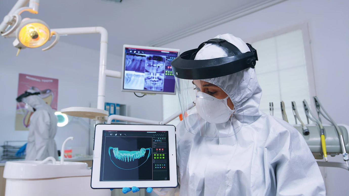
X-rays, also known as radiographs, are used in a variety of medical setups. X-rays are mostly used to diagnose tumors and bone injuries. This technology is used in dental clinics to examine patients’ teeth and jaw bones in detail. Dental X-rays are an important part of dental care because they aid in diagnosis and preventive care for your teeth.
What are the uses of X-rays?
- Radiographs, or dental X-rays, are used to diagnose dental infections. The dentist uses the X-ray results to detect infection symptoms before they become visible. This allows those conditions to be treated as early as possible before they worsen and necessitate more advanced treatment.
- The X-rays provide images of parts of your mouth that the dentist cannot see, creating a vivid picture of the differences in your soft tissues. X-rays of the teeth may reveal:
- Abscesses and cysts
- Bone loss.
- Decay below the fillings
- Tumors, both cancerous and non-cancerous.
- Decay between the teeth
- Developmental anomalies.
- Tooth and root positions
- Internal tooth problems or problems below the gumline
Early detection and treatment of dental problems can save you time, money, unnecessary discomfort, and your teeth.
What are the various types of X-rays?
There are four types of dental X-rays, each of which captures a different aspect of the teeth.
- Bitewing X-rays are used to detect cavities and periodontal disease by showing both the upper and lower teeth.
- Periapical X-rays show the entire tooth, from the root to the crown. This type of X-ray is used to look at a specific area of concern, such as a toothache.
- Occlusal X-rays show how the upper and lower teeth line up on the floor of the mouth. This type of X-ray is commonly used to monitor a child’s tooth development.
- Panoramic X-rays show all of the lower and upper teeth, as well as the jaw and sinus area. This X-ray is most commonly used for orthodontic treatment or dental implants, and it can also detect impacted teeth, jaw disorders, cysts, and tumors.
Are dental X-rays safe?
- While some radiation is present, the level of exposure is deemed safe for both adults and children. According to studies, radiographs used in dentistry account for only 2.5 percent of all radiographs used in medical fields. Furthermore, when the dentist uses an X-ray on you, your exposure is lower than when the dentist uses it to develop an image on film.
- It is also worth noting that dentists receive X-ray training, and your vital organs are always protected from unnecessary exposure. There is no need to be concerned about radiation exposure from dental X-rays.
- If you are pregnant, you should avoid having dental X-rays. Radiation is harmful to a developing foetus. Inform your dentist that you are pregnant during the examination so that he does not perform an x-ray on you.
How frequently should dental X-rays be performed?
- The need for dental X-rays is determined by each patient’s specific dental health needs. Based on a review of your medical and dental history, dental examination, signs and symptoms, age consideration, and disease risk, your dentist will recommend the necessary X-rays.
- For new patients, a full-mouth series of dental X-rays is recommended. Bite-wing X-rays are taken at recall (check-up) visits and are recommended once or twice a year to detect new dental problems.
To summarize, it is critical to get dental X-rays to diagnose and treat problems early, which can potentially save you money and unnecessary discomfort in the future and help keep your teeth healthy and strong for a lifetime.


